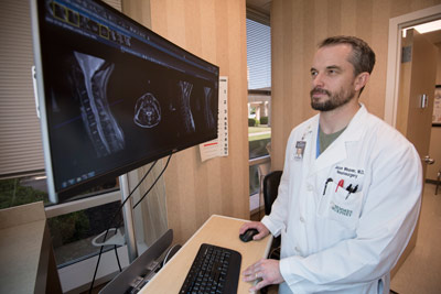
We treats many disorders of the spine, including:
Herniated Disc Symptoms
Symptoms vary greatly depending on the position of the herniated disc and the size of the herniation. If the herniated disc is not pressing on a nerve, the patient might experience spinal pain (cervical, lumbar, and thoracic) or no pain at all. If there is pressure on a nerve, there can be pain, numbness or weakness in the area of the body to which the nerve travels. Typically, a herniated disc is preceded by an episode of spinal pain (cervical, lumbar, and thoracic) or a long history of intermittent episodes of spinal pain.
- Lumbar spine (lower back) — Sciatica frequently results from a herniated disc in the lower back. Pressure on one or several nerves that contribute to the sciatic nerve can cause pain, burning, tingling, and numbness that radiates from the buttock into the leg and sometimes into the foot. Usually one side (left or right) is affected. This pain often is described as sharp and electric shock-like. It may be more severe with standing, walking or sitting. Along with leg pain, you may experience low back pain. Using the term radiculopathymay be more appropriate, rather than using the term sciatica, since all leg pain isn’t necessarily “sciatica.”
- Cervical spine (neck) — Symptoms may include dull or sharp pain in the neck or between the shoulder blades, pain that radiates down the arm to the hand or fingers, or numbness or tingling in the shoulder or arm. The pain may increase with certain positions or movements of the neck.
- Thoracic spine — Symptoms of a thoracic disc herniation can be comprised of posterior chest pain radiating around one or both sides of the rib cage. Such pain is usually triggered by physical exertion and can even be caused by taking a deep breath. Bands of numbness around the chest wall can also be present. Herniated discs of the thoracic spine are relative rare compared to cervical and lumbar disc herniations.
SURGICAL TREATMENT OPTIONS:
Artificial disc surgery (disc arthroplasty) includes surgical replacement of a diseased or herniated cervical or lumbar disc with an artificial disc designed to maintain spinal mobility. These usually consist of a plastic core between two metallic (usually titanium) plates that lock into the spine.
Patients with cervical disc herniation that require surgery most often undergo Anterior Cervical Discectomy with Fusion (ACDF). This procedure requires the surgeon to operate through the front of the neck and can be performed using many types of implants including anterior titanium metallic plates and screws or intra-discal implants not requiring anterior plating (low or no profile implants). These implants are made of titanium, plastic, or a combination
Other less commonly used procedures include anterior and posterior microdiscectomy usually without fusion. Some cases of extensive cervical stenosis require de-compressive posterior laminectomy or laminoplasty augmented often by instrumented posterior cervical fusions (titanium rods, screws, plates). Alternatively, even these types of cases can be performed from the front of the neck and the surgery is called a corpectomy, with instrumented or metallic cage fusion.
Patients with lumbar disc herniation that require surgery are most commonly treated with micro-discectomy or other minimally invasive techniques to simply remove the herniated disc without destabilizing the spine. The indications to perform this procedure or others can be confusing and requires clear communication between patient and surgeon.
Scoliosis
Scoliosis is an abnormal lateral curvature of the spine. It is most often diagnosed in childhood or early adolescence. The spine’s normal curves occur at the cervical, thoracic, and lumbar regions in the so-called “sagittal” plane. These natural curves position the head over the pelvis and work as shock absorbers to distribute mechanical stress during movement. Scoliosis is often defined as spinal curvature in the “coronal” (frontal) plane. While the degree of curvature is measured on the coronal plane, scoliosis is actually a more complex, three-dimensional problem which involves the following planes: Coronal plane, Sagittal plane, Axial plane.
The coronal plane is a vertical plane from head to foot and parallel to the shoulders, dividing the body into anterior (front) and posterior (back) sections. The sagittal plane divides the body into right and left halves. The axial plane is parallel to the plane of the ground and at right angles to the coronal and sagittal planes.
Symptoms/Signs: There are several signs that may indicate the possibility of scoliosis. If you notice one or more of the following signs, you should schedule an appointment with a doctor.
- Shoulders are uneven – one or both shoulder blades may stick out
- Head is not centered directly above the pelvis
- One or both hips are raised or unusually high
- Rib cages are at different heights
- Waist is uneven
- The appearance or texture of the skin overlying the spine changes (dimples, hairy patches, color abnormalities)
- The entire body leans to one side
Diagnosis: Scoliosis is usually confirmed through a physical examination, an x-ray, spinal radiograph, CT scan or MRI. The curve is measured by the Cobb Method and is diagnosed in terms of severity by the number of degrees. A positive diagnosis of scoliosis is made based on a coronal curvature measured on a posterior-anterior radiograph of greater than 10 degrees. In general, a curve is considered significant if it is greater than 25 to 30 degrees. Curves exceeding 45 to 50 degrees are considered severe and often require more aggressive treatment.
Treatment: When there is a confirmed diagnosis of scoliosis, there are several issues to assess that can help determine treatment options:
- Spinal maturity – is the patient’s spine still growing and changing?
- Degree and extent of curvature – how severe is the curve and how does it affect the patient’s lifestyle?
- Location of curve – according to some experts, thoracic curves are more likely to progress than curves in other regions of the spine.
- Possibility of curve progression – patients who have large curves prior to their adolescent growth spurts are more likely to experience curve progression.
After these variables are assessed, treatment options can include observation, bracing, or surgery depending on the recommendations of your doctor.
Osteoarthritis
Osteoarthritis, in general, is the most common type of arthritis and affects middle-aged or older people most frequently. It can cause a breakdown of cartilage in joints and occur in almost any joint in the body. It most commonly affects the hips, knees, hands, lower back and neck. Cartilage is a firm, rubbery material that covers the ends of bones in normal joints. It serves as a kind of “shock absorber,” helping to reduce friction in the joints.
When osteoarthritis affects the spine, it is known as spondylosis. Spondylosis is a degenerative disorder that can cause loss of normal spinal structure and function. Although aging is the primary cause, the location and rate of degeneration varies per person. Spondylosis can affect the cervical, thoracic and/or lumbar regions of the spine, with involvement of the intervertebral discs and facet joints. This can lead to disc degeneration,bone spurs, pinched nerves, and an enlargement or overgrowth of bone that narrows the central and nerve root canals, causing impaired function and pain.
When spondylosis affects the lumbar spine, several vertebrae usually are involved. Because the lumbar spine carries most of the body’s weight, activity or periods of inactivity can both trigger symptoms. Specific movements, sitting for prolonged periods of time, and lifting and bending all may increase pain.
When spondylosis worsens, a patient may develop spinal stenosis — a narrowing of spaces in the spine that results in pressure on the spinal cord and/or nerve roots. The narrowing can affect a small or large area of the spine. Pressure on the upper part of the spinal cord may produce pain or numbness in the shoulders and arms. Pressure on the lower part of the spinal cord or on nerve roots branching out from that area may cause pain or numbness in the legs.
Degenerative spondylolisthesis (slippage of one vertebra over another) is caused by osteoarthritis of the facet joints. Most commonly, it involves the L4 slipping over the L5 vertebra. It most frequently affects people age 50 and older. Symptoms may include pain in the low back, thighs, and/or legs, muscle spasms, weakness, and/or tight hamstring muscles.
Symptoms
- Pain and stiffness in the neck or low back
- Pain that radiates into the shoulder or down the arm
- Weakness or numbness in one or both arms
- Pain or morning stiffness that lasts for about 30 minutes due to inactivity
- Pain that worsens throughout the day due to activity
- Limitation of motion
Causes
While the cause of osteoarthritis is unknown, the following factors may increase the risk of developing the condition:
- Age
- Heredity
- Being overweight
- Joint injury
- Nerve injury
- Repeated overuse of specific joints
- Lack of physical activity
Diagnosis
A diagnosis usually can be made based on specific symptoms, a thorough physical examination and x-ray results. On occasion, magnetic resonance imaging (MRI) may be ordered to determine the extent of damage in the spine. MRI can reveal damaged cartilage, loss of joint space or bone spurs.
Nonsurgical Treatment
- Anti-inflammatory medications to reduce swelling and pain, and analgesics to relieve pain. Most pain can be treated with nonprescription medications, but if pain is severe or persistent, your doctor may recommend prescription medications.
- Epidural injections of cortisone may be prescribed to help reduce swelling. This treatment is not recommended repeatedly and usually provides only temporary pain relief.
- Physical therapy and/or prescribed exercises may help stabilize your spine, build your endurance and increase your flexibility. Therapy may help you resume your normal lifestyle and activities. Yoga may be effective for some people in helping to manage symptoms.
- Maintaining a proper weight is crucial to effective management of osteoarthritis. Being overweight is a risk factor for osteoarthritis.
Surgical treatment for spondylosis is uncommon, unless the condition has led to severe spinal stenosis. Surgery may be recommended if conservative treatment options, such as physical therapy and medications, do not reduce or end the pain altogether, and if the pain greatly impairs the person’s daily functions. As with any surgery, a patient’s age, overall health and other issues are taken into consideration when surgery is considered.
Information courtesy of the American Association of Neurological Surgeons
Publications
- Domagoj Gajski, Alicia R. Dennis, Kenan I. Arnautović: Surgical anatomy of microsurgical 3-level anterior cervical discectomy and fusion C4–C7, BJBMS, DOI: https://dx.doi.org/10.17305/bjbms.2020.4895. Published: 20 June 2020
- Mullins, J; Pojskic, M; Boop, FA; Arnautovic, KI, Retrospective single-surgeon study of 1,123 consecutive cases of anterior cervical discectomy and fusion: A comparison of clinic, outcome parameters, and costs between outpatient and inpatient surgery groups with a literature review, Journal of Neurosurgery Spine, June 2018, 630-641.
- Muzevic, D; Spavski B; Boop FA; Arnautovic, KI, Anterior cervical discectomy with instrumented allograft fusion: Lordosis restoration and comparison of functional outcomes among patients of different age groups, World Neurosurgery October: pii: 21878-8750 (17) 31662-5. doi 10.1016/j.wneu.2017.09.146, e1049-1062, 2016
- Arnautovic, KI, Olinger, R.G., Anterior Cervical Diskectomy, Drainage and Non-instrumented Cortico-Cancellous Allograft Fusion: A Treatment Option for Ventral Cervical Spinal Epidural Abscess. Medical Archives 66: 194-197, 2012.
- Arnautovic, KI, New from the Journals, In the Eye of the Beholder: Preferences of Patients, Family Physicians and Surgeons for Lumbar Spine Surgery. Spineline 11:30-31, 2010
- Al-Mefty R, Arnautovic KI, Webber BL., Multilevel bilateral calcified thoracic spinal synovial cysts. Journal of Neurosurgery (Spine) 8: 473-477. 2008.
- Pait TG, Castro I, Arnautovic KI, Borba LAB. Compound osteosynthesis in the thoracic spine for treatment of vertebral metastases. Technical report. Arquivos Neuro-Psiquiatria 58: 52-56, 2000.
- Pait TG, Al-Mefty O, Boop FA, Arnautovic KI, Rahman S, Ceola W. Inside-outside technique for occipital and posterior cervical spine instrumentation and stabilization: Preliminary results. Journal of Neurosurgery (Spine) 90: 1-8, 1999 (Cover Article)
- Pait T G, Arnautovic KI, Borba L A B. Microsurgical anatomy of the atlantoaxial region. Perspectives in Neurological Surgery 7: 91-98, 1997
- Pait T G, Killefer J A, Arnautovic KI. Surgical anatomy of the anterior cervical spine: The disc space, vertebral artery, and associated bony structures. Neurosurgery 39: 769-777, 1996.
- Kadic N, Arnautovic KI. Percutaneous lumbar diskectomy guided by computer tomography. Advantages and disadvantages. Proceedings of the 2nd Course on Percutaneous Lumbar Diskectomy, Zagreb, Croatia: 85-92, 1991.
- Arnautovic KI. Spinal cord injury without radiological abnormality (SCIWORA): the incidence, pathology, clinical observation and treatment. Chirurgia Neurologica 1: 40-54, 1990.
- Kadic N, Arnautovic KI, Percutaneous lumbar diskectomy using CT scan as a radiological guide. Chirurgia Neurologica 1: 19-22, 1990.
- Arnautovic KI, Konjhodzic F. A conservative treatment of the isolated spinal cord injuries. Proceedings of the International Symposium on Spine and Spinal Cord Diseases and Injuries, Varazdinske Toplice, Croatia: 87-97, 1989.
- Kadic N, Custovic K, Arnautovic KI. Operative treatment of the cervical disc lesions using anterior approach without intervertebral fusion. Medicinski Arhiv 42: 281-285, 1988.
- Custovic K, Skljarevski V, Jurisic, Arnautovic KI. Characteristics of cervical spinal injuries associated with head injuries. Proceedings of the 11th World Congress of the International Association for Accident and Traffic Medicine, Dubrovnik, Croatia: 333-337, 1998.

